.png)
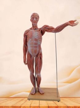
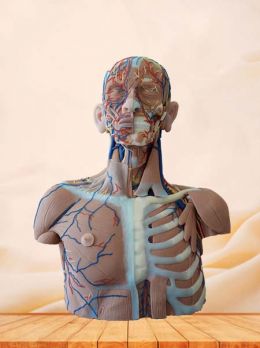
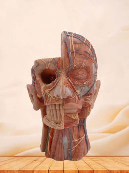
Combination of virtual anatomy and real anatomical specimen
Meiwo Science is a large manufacturer appointed by National Ministry of Education and one of the large models producing factory in China, Specialized in plastination technique, soft silicone anatomy models, high simulation anatomy models and relative production, The main products are Plastination for animals, soft silicone anatomy models, high simulation anatomy models, human & animal anatomy softwares, embedded specimens and biological microscope slides, etc.
Meiwo factory lab do deep research and development for plastination and silicone anatomy model and do professional plastination from step by step, for example, embalming and fixation, anatomical dissection, removal water and fatty, forced impregnation, position, Harden, etc. We are glad to teach you the more knowledge about plastination, silicone anatomy models, anatomy softwares and also can see our real animal plastinated specimens, silicone anatomy models, anatomy softwares...
International medical exhibition in Moscow, Russia is held 8 to 12 DEC,2014. Exhibition area of 45,000 square meters, an...
MEIWO take part in the FIME on 6 to 8 August, 2014 in Miami Beach exhibition hall in USA.
HOSPITALAR is held 21st to 24th May 2014 in Sao Paulo, Brazil. Meiwo attend this exhibition and show our whole body post...
The production of prepared microscope slides set is completed, and final packaging and inspection are being performed. R...
On Mar 26th 2015,one of close and old friends from Saudi Arabia finally to see me and I am so happy to see him. Mr Joh...
On July 26th 2015 Meiwo science deliver the 3 cartons human anatomy models including the human skeleton models, heart an...
The interesting thing about human anatomy: the number of human bones is different in infants and adults.
The new animal plastination museum for Agriculture and Animal Husbandry University will be finished by meiwo science. Al...
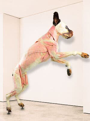
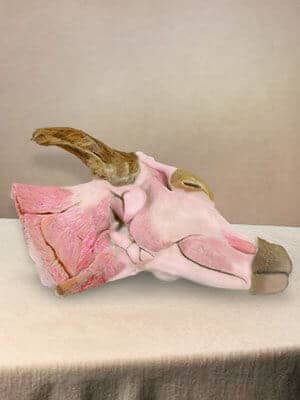
A series of plastinated animals finished by Meiwo, including plastinated cat, plastinated dog, plastinated pig, plastina...
 English
 Spanish