
Combination of virtual anatomy and real anatomical specimen
Meiwo Science is a large manufacturer appointed by National Ministry of Education and one of the large models producing factory in China, Specialized in plastination technique, soft silicone anatomy models, high simulation anatomy models and relative production, The main products are Plastination for animals, soft silicone anatomy models, high simulation anatomy models, human & animal anatomy softwares, embedded specimens and biological microscope slides, etc.
Meiwo factory lab do deep research and development for plastination and silicone anatomy model and do professional plastination from step by step, for example, embalming and fixation, anatomical dissection, removal water and fatty, forced impregnation, position, Harden, etc. We are glad to teach you the more knowledge about plastination, silicone anatomy models, anatomy softwares and also can see our real animal plastinated specimens, silicone anatomy models, anatomy softwares...
According to the academic exchange plan of the CSAS in 2022, sponsored by the Nursing Anatomy Branch of the CSAS and undertaken by Hexi College in Zhangye City, Gansu Province, The 17th Annual Conference of Nursing Anatomy, co-organized by Gansu Anatomy Society, Guangdong Anatomy Society and Anatomy Research Magazine, will be held in Hexi College, Zhangye City, Gansu Province from July 17 to 21, 2022.
Due to the proposal of the concept of "meta-universe" and the development of artificial intelligence technology, the virtualization of real human body and the reality of virtual human body are accelerating, and the informatization of human body structure and function has ushered in an important period of strategic opportunity. In order to further promote the research and development of digital human body and related products in China, The CSAS decides to hold the Digital Human Science and Technology Frontier and Application Forum in Jinan city from 20th to 22th, May in 2022 . Considering the current situation of COVID-19 prevention and control in China, the meeting was conducted in a combination of offline and online methods
The new morphology virtual anatomy software for medical college as an important part of microbiology teaching has been finshed and all the equipments have also been installed.
Heart anatomy is a core and challenging topic in human anatomy, physiology, pathology, and clinical medicine. The native human heart is relatively small, with intricate internal chambers, delicate valves, and complex vascular branches and spatial relationships. Relying solely on textbook images and text descriptions is insufficient for learners to develop a three-dimensional understanding and cannot meet the needs of refined teaching. High simulatiom magnified heart anatomy models, based on the real human heart and scaled up proportionally, achieve a high degree of realism in morphology, structure, and tissue texture. This effectively compensates for the shortcomings of traditional teaching methods and possesses multiple teaching values, including basic anatomy instruction, explanation of physiological functions, pathological correlation analysis, and connection to clinical applications. It is an indispensable core teaching tool in medical-related majors. These high simulation heart anatomy models strictly adhere to real human cardiac anatomical data, accurately reproducing all core and minute structures, including the left and right atria, left and right ventricles, atrial septum, interventricular septum, various valves (mitral, tricuspid, aortic, and pulmonary valves), papillary muscles, chordae tendineae, main and branch coronary arteries, coronary sinuses, and remnants of the foramen ovale. Meanwhile, the model accurately reproduces the differences in myocardial wall...
High simulation anatomical models of uterus, ovaries, and fallopian tubes are core teaching aids in medical education, providing intuitive, concrete, and interactive representations. They are particularly suitable for teaching and training in obstetrics and gynecology, reproductive medicine, nursing, midwifery, and maternal and child health, serving applications in theoretical instruction, skills training, clinical communication, and public education. Anatomy itself is abstract and three-dimensional. Relying solely on textbooks, atlases, and two-dimensional images makes it difficult for learners to develop a complete understanding of organ morphology, positional relationships, and adjacent structures. These high simulation anatomical models of uterus, ovaries, and fallopian tubes are meticulously crafted according to standard human anatomy proportions and morphology, fully presenting the uterus’s shape, uterine wall layers, and uterine cavity morphology; the ovary’s size, cortical and medullary structure, and follicular development stages; and the fallopian tubes’ interstitial portion, isthmus, ampulla, fimbriae, and their connections to the uterus, ovaries, and pelvic wall. Learners can intuitively understand the three-dimensional spatial position of pelvic organs and the role of uterine ligaments in fixing the uterus’s position through touch, observation, and disassembly of the models. This understanding is fundamental to comprehending the physiological and pathological mechanisms of the reproductive system. Highly realistic anatomical...
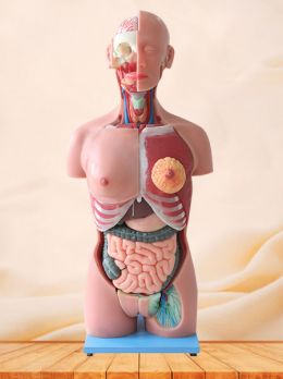
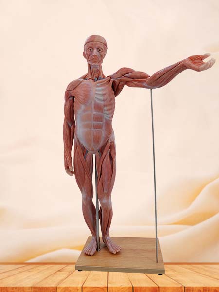
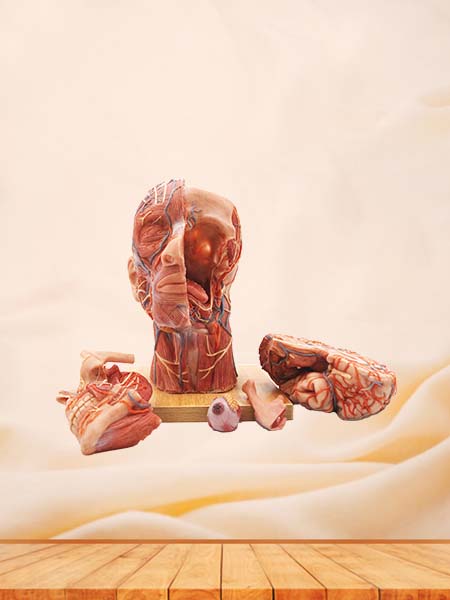
Human torso model is a meticulously crafted model based on a simulation of the existing human torso. It features detailed anatomical dissections for easy observation, making it a highly realistic human model suitable for teaching and research. Hongyu meticulously dissects each organ of the human torso, using different colors for different organs or replacing diseased organs with normal ones in the same location. This allows for intuitive and visual education and demonstration. Furthermore, using modern high-tech methods such as ultrasound, X-rays, and CT scans, it allows for detailed simulation and research of the normal and pathological structures and conditions of the internal organs of the human torso. It has the advantages of being easy to observe, convenient for teaching, and beneficial for research. Meiwo human torso model comprises 27 parts, displaying the location of internal organs and the anatomical morphology and structure of the head, representing the respiratory, digestive, and urinary systems. The right half of the skull shows the skull, masseter muscle, temporalis muscle, and other structures. The eyeballs are visible within the orbits. A sagittal section of the head and neck is shown, revealing the cranial cavity containing the right hemisphere of the brain. The ventral surface of...
With the modernization of agriculture and animal husbandry, agricultural and animal husbandry colleges are increasingly demanding higher standards in talent cultivation. Beyond basic theoretical knowledge, students’ practical skills have become a crucial area of focus. To help students better understand professional knowledge such as animal anatomy and disease control, animal specimens, as a unique teaching resource, have become an indispensable tool in agricultural and animal husbandry disciplines. Animal specimens not only provide students with a direct learning experience but also play a vital role in enhancing practical skills and promoting educational innovation. Animal specimens play an irreplaceable role in agricultural and animal husbandry teaching. Compared to pictures, illustrations, or models in textbooks, specimens offer students a more realistic and concrete learning experience. Whether it’s a cattle, sheep, or other common livestock specimen, students can gain a deeper understanding of the functions of various animal systems and their interrelationships by observing the actual structure of the specimen. Through observing animal specimens, students can clearly see the morphology, structure, and physiological functions of different organs within the animal’s body. For example, through a cattle digestive system specimen, students can not only see the location of organs such as the stomach and intestines...
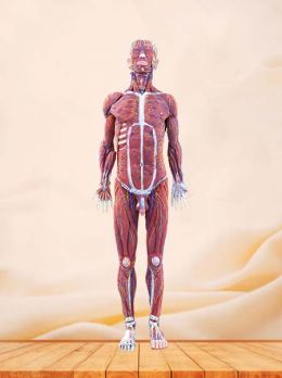
In the development of medical education, the innovation of teaching tools has always been a crucial force driving the improvement of educational quality. From early simple skeletal specimens to today’s advanced high-fidelity human anatomical models, the evolution of these teaching aids has profoundly influenced the methods and effectiveness of medical teaching. High-fidelity human anatomical models, with their unique advantages, are gradually becoming a new favorite in the field of medical education, bringing unprecedented teaching experiences to teachers and students. Meiwo Science’s high-fidelity human anatomical models use environmentally friendly, food-grade soft silicone material, completely free of harmful heavy metals such as lead, mercury, cadmium, and hexavalent chromium. These models possess excellent stability and durability, and are not prone to aging or deformation even after long-term use. The non-toxic and odorless material ensures the health and safety of users, avoiding potential safety hazards and ethical issues associated with traditional anatomy. Furthermore, the soft silicone material has excellent softness and elasticity, realistically simulating the structure of human muscles, skin, and internal organs. Even after repeated bending and washing, the models retain their original shape, exhibiting extremely high durability and economy, making them suitable for long-term, repeated use. Meiwo Science anatomical models not only...
In the current era of continuous development in medical education and advancements in technology, virtual anatomy specimen databases, as an emerging digital tool, are deeply integrated into the teaching and research systems of medical schools, bringing about a revolutionary change in medical talent cultivation. With its rich digital resources and unique functions, it plays an irreplaceable and vital role in medical schools. Traditional medical anatomy teaching relies on physical specimens, which suffers from limited specimen quantity, high loss, and limited morphological diversity, making it difficult to meet diverse teaching needs. Virtual anatomy specimen databases, possessing massive amounts of high-definition, multi-angle virtual anatomical images and 3D models, break these limitations. In classroom teaching, teachers can use the database to present complex human structures to students in a vivid and intuitive way. For example, when explaining the structure and function of the heart, a 3D model can comprehensively display the internal structure of the heart, including the direction of myocardial fibers and the opening and closing mechanism of valves. Students can more clearly and accurately understand the working principle of the heart, greatly improving the efficiency of knowledge transfer compared to traditional two-dimensional images and text descriptions. Furthermore, virtual anatomy specimen databases...
 English