
Combination of virtual anatomy and real anatomical specimen
Meiwo Science is a large manufacturer appointed by National Ministry of Education and one of the large models producing factory in China, Specialized in plastination technique, soft silicone anatomy models, high simulation anatomy models and relative production, The main products are Plastination for animals, soft silicone anatomy models, high simulation anatomy models, human & animal anatomy softwares, embedded specimens and biological microscope slides, etc.
Meiwo factory lab do deep research and development for plastination and silicone anatomy model and do professional plastination from step by step, for example, embalming and fixation, anatomical dissection, removal water and fatty, forced impregnation, position, Harden, etc. We are glad to teach you the more knowledge about plastination, silicone anatomy models, anatomy softwares and also can see our real animal plastinated specimens, silicone anatomy models, anatomy softwares...
According to the academic exchange plan of the CSAS in 2022, sponsored by the Nursing Anatomy Branch of the CSAS and undertaken by Hexi College in Zhangye City, Gansu Province, The 17th Annual Conference of Nursing Anatomy, co-organized by Gansu Anatomy Society, Guangdong Anatomy Society and Anatomy Research Magazine, will be held in Hexi College, Zhangye City, Gansu Province from July 17 to 21, 2022.
Due to the proposal of the concept of "meta-universe" and the development of artificial intelligence technology, the virtualization of real human body and the reality of virtual human body are accelerating, and the informatization of human body structure and function has ushered in an important period of strategic opportunity. In order to further promote the research and development of digital human body and related products in China, The CSAS decides to hold the Digital Human Science and Technology Frontier and Application Forum in Jinan city from 20th to 22th, May in 2022 . Considering the current situation of COVID-19 prevention and control in China, the meeting was conducted in a combination of offline and online methods
The new morphology virtual anatomy software for medical college as an important part of microbiology teaching has been finshed and all the equipments have also been installed.
On Dec 8th 2014 the new year is coming soon, Mr Christian from Canada do the cooperation with us, and he likes our products very much. and do the samples. when he received the samples. He said: "Amy, your products is very amazing and now we will place another order. " We feel very proud of our unique products.
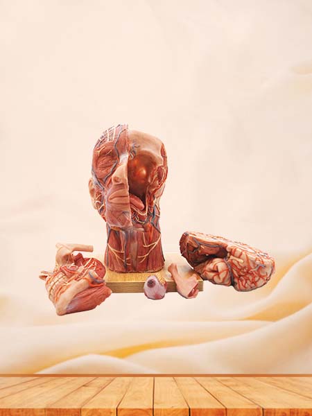
Meiwo new anatomy model_anterior, middle and posterior of neck for medical education has been finished, it was made of soft silicone rubber, nontoxic, odorless, durable, fall and crash resistant.
A series of plastinated organs of cow for veterinary education have been finished by professional anatomy teachers and the veterinary anatomy professor has spoken highly of our plastinated organs of cow.A series of plastinated organs of cow for veterinary education have been finished by professional anatomy teachers and the veterinary anatomy professor has spoken highly of our plastinated organs of cow.
Sectional anatomy is a discipline that combines anatomy and radiology. Through anatomical analysis and study of human body tomography images, the internal structure of human body can be accurately located and described. In the teaching of human anatomy, sectional anatomy has an important position and significance.
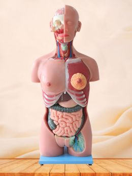
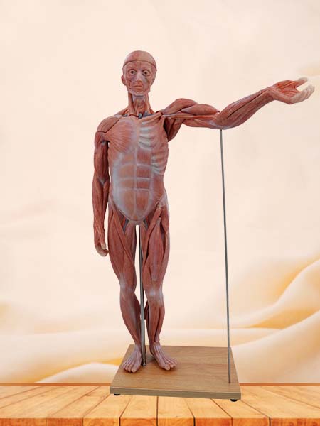
Do you know about soft silicone anatomy model for medical education? The soft silicone anatomy model is made of environmental silicone rubber. The silicone rubber and its auxiliary chemical products used in medical soft silicone anatomical models have passed ROHS certification to ensure the stability and durability of the models. The soft silicone anatomical models have passed FDA certification, non-toxic and tasteless, accurate structure, realistic shape, can be repeatedly bent, durable, easy to disassemble and clean and so on.
With the development of The Times, more and more medical models have come into people's attention. Besides being used in basic medical anatomy experiments and clinical surgery teaching, medical models are also used in popular science exhibitions such as museums, art galleries and popular science venues. Besides being used in basic medical anatomy experiments and clinical surgery teaching, medical models are also used in popular science exhibitions such as museums, art galleries and popular science venues. These models help people learn some of the necessary first aid techniques and understand the basic structure of the human body.
 English